Atlee_2_confocal_2013_600
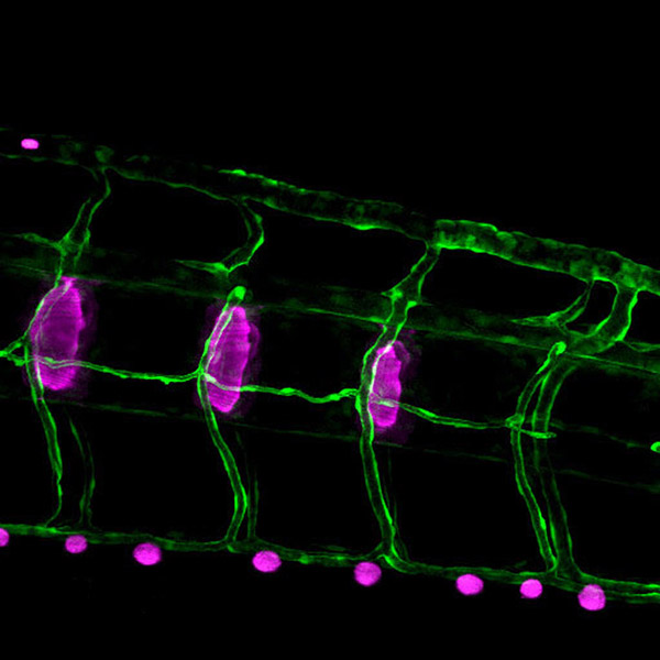
Maximum intensity projection of vertebral body. Blood vessels (gfp, shown in green) and mineralized bone matrix (shown in magenta. Field width: 0.42 mm.
Image by AtLee Watson and Melissa Pickett, confocal microscopy class of 2013.