Marlee_Trandel_01_600
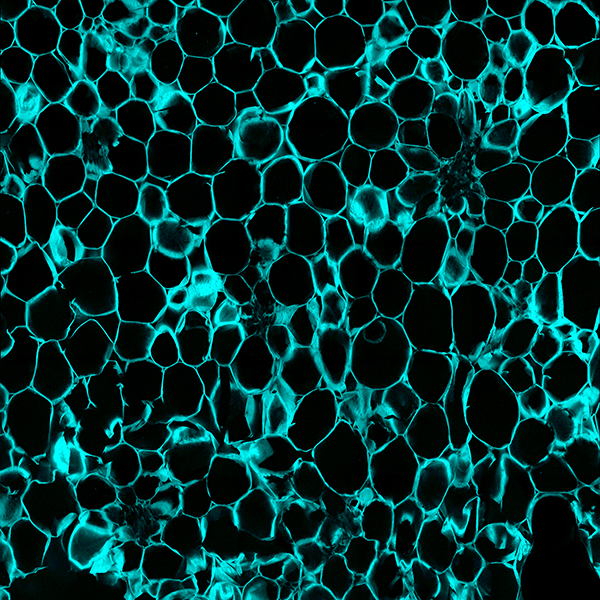
Watermelon placental (heart) tissue is displayed by use of calcofluor white dye. This confocal micrograph was taken to determine the number of cells and obtain the average surface area of watermelon cell tissue in control fruit compared to fruit with an internal defect known as hollow heart. Hollow heart is most dominantly found in non-grafted, seedless watermelon. The cause of the defect is still unknown, but is mostly correlated to field issues such as watermelon pollination and water stress. Watermelon cultivars with increases in tissue firmness or tissue density (i.e., the number of cells per unit area) are also known to be less susceptible to this serious internal fruit disorder. This micrograph was obtained to better correlate tissue density to hollow heart susceptibility in a seedless cultivar. Tile scan on Zeiss LSM 880. Image taken by Marlee Trandel. Research Lab: Dr. Penelope Perkins-Veazie, Horticultural Sciences.