- This event has passed.
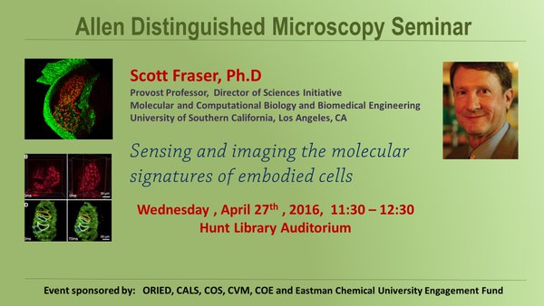
Scott Fraser: Sensing and imaging the molecular signatures of embodied cells
April 27, 2016 @ 11:30 am - 12:30 pm

Allen Distinguished Microscopy Seminar
Sensing and imaging the molecular signatures of embodied cells
Scott Fraser, Ph.D, Provost Professor, Director of Sciences Initiative
Molecular and Computational Biology and Biomedical Engineering
University of Southern California, Los Angeles, CA
Sponsored By:
ORIED, CALS, COS, CVM, COE & Eastman Chemical University Engagement Fund
Abstract: The challenge of modern embryology is to draw upon the growing body of high-throughput molecular data to better understand the cellular and molecular events underlying embryonic development. This wealth of data now presents the challenge of integrating a working knowledge of how these molecular components, often present at vanishingly small concentrations, generate reliable patterns of cell migration and cell differentiation. In typical cell biology approaches, cultures of isolated cells have been used to reveal mechanism. What is needed to understand development is to carry out studies on cells in their normal context interacting with other cells and signals in the intact embryo.
Imaging techniques are challenged by major tradeoffs between spatial resolution, temporal resolution, and the limited photon budget. We are attempting to advance this tradeoff by constructing faster and more efficient light sheet microscopes that maintain subcellular resolution. Our two-photon light-sheet microscope combines the deep penetration of two-photon microscopy and the speed of light sheet microscopy to generate images with more than ten-fold improved imaging speed and sensitivity. As with other light sheet technologies, the collection of an entire 2-D optical section in parallel offers dramatically speeds acquisition rates. Two-photon light sheet microscopy is far less subject to light scattering, permitting subcellular resolution to be maintained far better than conventional light sheet microscopes. The combination of attributes permits cell and molecular imaging with sufficient speed and resolution to generate unambiguous tracing of cells and signals in intact systems.
Multispectral imaging offers the chance of asking multiple questions of the same embodied cells. Multiplex analyses permit the variance and the “noise” in a system to be exploited by asking about the analytes that co-vary with a selected gene product. These approaches allow straightforward prediction of draft gene regulatory networks.
In parallel, label-free approaches offer an important approach for molecular sensing, but the low concentrations and low sensitivity of the techniques can make single cell approaches challenging. We have refined a new technology for enhancing these signals and have achieved gains that make the analysis of single cells possible.