Planchart_lab_01_600
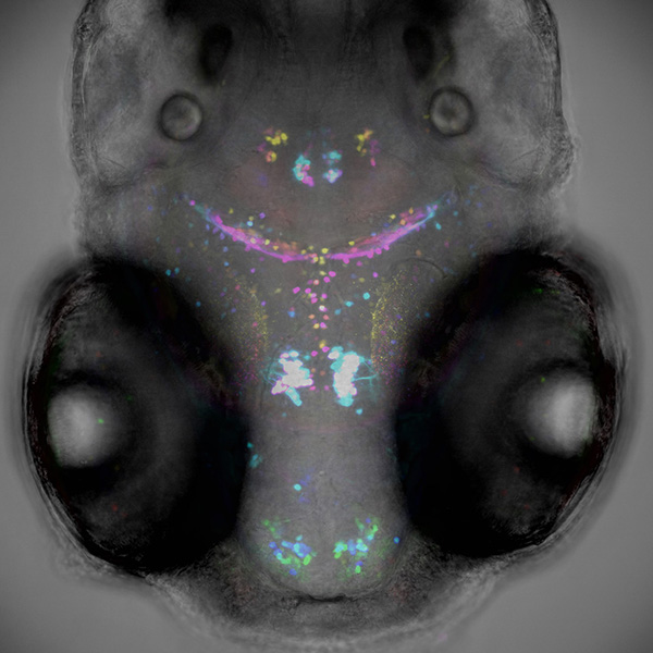
Dopaminergic neurons in the brain of a live 5-day old zebrafish. Maximum intensity projection, neurons are colored based on their depth in the brain. The fluorescence image is merged with a transmitted light image that was captured simultaneously. This fish is utilized by a toxicology lab to visualize how developmental exposure to environmental toxicants can affect the number and/or location of this specific type of neuron. Field width 0.6 mm. Image by Elizabeth Cook. Research groups: Drs. Antonio Planchart and Carolyn Mattingly, Biological Sciences, Center for Human Health and the Environment (CHHE).